Otorrhea, the medical term for any drainage from the ear, can range from harmless clear fluid to foul-smelling pus or even blood-tinged discharge. While it often signals an underlying ear condition, timely evaluation and tailored treatment usually lead to excellent outcomes. This article offers a streamlined overview of otorrhea—its types, causes, clinical presentation, diagnostic approach, management strategies, potential complications, and preventive measures.
1. What Is Otorrhea?
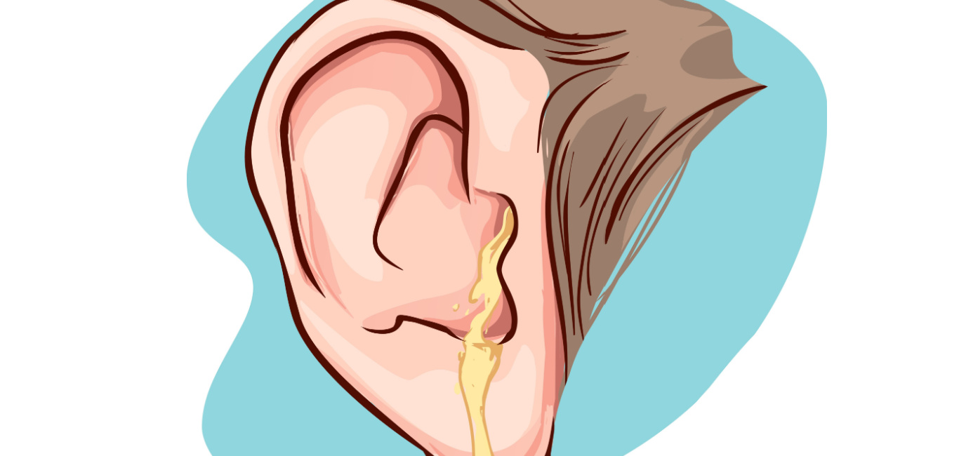
Otorrhea refers to fluid that exits the external auditory canal or middle ear via a perforated eardrum. The discharge’s color and consistency provide clues about its origin and severity.
2. Classification by Character
-
Serous: Thin, watery, usually non-infectious.
-
Purulent: Thick, yellow-green, often bacterial.
-
Mucoid: Sticky, mucus-like, typical of chronic inflammation.
-
Serosanguineous: Mixed with blood, may follow trauma or surgery.
-
Clear: Odorless and non-mucoid, often benign.
3. Common Causes
-
Acute Otitis Media with Perforation
Sudden eardrum rupture from middle-ear infection lets purulent fluid drain. -
Otitis Externa (“Swimmer’s Ear”)
Infection or inflammation of the ear canal leads to itchiness and discharge. -
Chronic Suppurative Otitis Media
Persistent or recurrent drainage lasting more than six weeks. -
Cholesteatoma
Abnormal skin growth in the middle ear that erodes bone and produces discharge. -
Trauma or Surgery
Skull-base fractures or ear-tube procedures can cause blood-tinged or clear leaks. -
Foreign Bodies
Objects in the ear canal may provoke irritation and drainage.
4. How Patients Present
-
Discharge: Variable odor and consistency—from clear to foul-smelling.
-
Ear Pain: Often sharp or throbbing, especially with acute infections.
-
Hearing Loss: Conductive loss from fluid blockage or membrane perforation.
-
Associated Symptoms: Itching, tinnitus, fullness, or occasional dizziness.
5. Diagnostic Approach
-
History
-
Duration: Acute (<6 weeks) vs. chronic (≥6 weeks)
-
Precipitating factors: Swimming, trauma, prior surgeries
-
Symptoms: Pain, fever, hearing changes
-
-
Physical Exam
-
Otoscopy: Assess eardrum integrity, canal inflammation, discharge character
-
-
Additional Tests (as needed)
-
Microbial culture of discharge for treatment-resistant cases
-
Audiometry to quantify hearing impairment
-
CT or MRI if cholesteatoma, mastoiditis, or skull-base fracture is suspected
-
6. Treatment Strategies
Medical
-
Topical Therapy
-
Antibiotic or antifungal ear drops
-
Gentle aural toilet (suctioning and cleaning)
-
-
Systemic Therapy
-
Oral antibiotics for middle-ear infections with systemic signs
-
Analgesics for pain relief
-
Surgical
-
Tympanoplasty
-
Repair of chronic eardrum perforations
-
-
Mastoidectomy
-
Removal of infected mastoid air cells in cases of mastoiditis or cholesteatoma
-
-
Ear-Tube Revision or Removal
-
Address persistent otorrhea in patients with tympanostomy tubes
-
7. Potential Complications
-
Permanent Hearing Loss: From ossicular chain damage or chronic inflammation.
-
Cholesteatoma Formation: Leading to bone erosion and more serious infection.
-
Intracranial Spread: Rare but severe, including abscess or meningitis.
8. Prevention and Patient Education
-
Protect Ears from Water: Use ear plugs or coated cotton during swimming.
-
Avoid Cotton-Tip Swabs: Can injure canal skin or eardrum.
-
Prompt Treatment of Ear Symptoms: Early care for ear pain or discharge to prevent chronicity.
9. Conclusion
Otorrhea is a hallmark symptom of various ear disorders. A structured evaluation—focusing on discharge characteristics, patient history, and targeted investigations—enables precise diagnosis. From topical drops and systemic antibiotics to surgical repair, management is tailored to the underlying cause. With appropriate treatment and preventive measures, most individuals fully recover with preserved hearing and minimal risk of complications.